When the world starts spinning: Understanding vertigo and tinnitus
July 7, 2026

Approximately 90% of vertigo cases can be diagnosed through careful history-taking, helping distinguish peripheral, central and other underlying causes.
Balance is the interaction between the vestibular system, which involves the ear; the ocular system, which involves the eyes; proprioception, which involves joint sensors; and the central nervous system, which integrates all these signals.
The semicircular canals are responsible for detecting angular rotational movement. The central part, where the saccule and utricle are located, detects linear movement, including horizontal and vertical motion. The cochlea is responsible for hearing. The vestibule and semicircular canals function as the sensory organs for balance. “We can recognize basically roll, pitch and yaw. When combined, that is what our vestibular system and central nervous system can detect. The utricle detects movement in the horizontal plane, meaning front and back movement. The saccule detects movement in the vertical plane, meaning up and down movement. “If you know you are in a lift moving up and down, that is because your saccule is working normally. If you are in a car and you know that you are moving forward, then your utricle is working normally,” explains Dr Shailendra Sivalingam (picture below), Consultant ENT, Head and Neck Surgeon, Base of Skull Surgeon and Neuro-Otologist at the ‘Modern Diagnostics and Advanced Therapeutics in ENT Care’ symposium by Sunway Medical Center, Sunway City. The semicircular canals detect rotational movement. The system is paired, meaning one canal on the right corresponds with another canal on the left that lies in the same plane. It functions through what is known as a push-and-pull relationship. “For example, if I turn to my left, the signal from the canal on the left will increase, while the signal from the canal on the right will decrease. This allows you to maintain movement. Patients with dysfunction of these canals develop vertigo because there is a mismatch of signals coming in.” Eye movement is also involved in maintaining balance through eye tracking. For example, if you extend your hand, look at your thumb, and turn your head left and right, the ability to maintain your gaze on your thumb is due to the lateral semicircular canal. Each lateral semicircular canal innervates the medial rectus muscle on the same side and the lateral rectus muscle on the opposite side, allowing coordinated eye tracking. 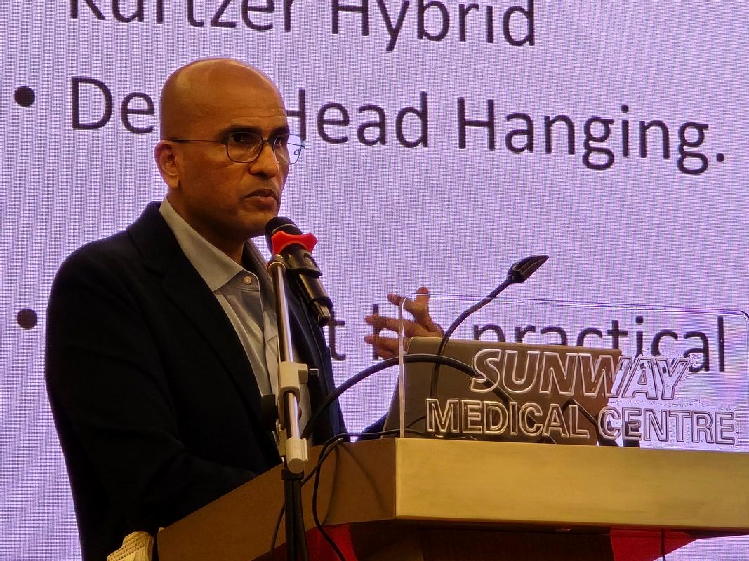
Fundamental concept
Proper balance is achieved through the harmonious interaction between the vestibulo-ocular reflex (VOR) and the vestibulo-spinal reflex. “The vestibulo-spinal reflex is demonstrated when you stand straight and close your eyes. The fact that you can remain standing and not fall backwards means you have a normal vestibulo-spinal reflex. All of this is controlled and managed by the higher centers in the cerebellum.” Causes of dizziness The causes of dizziness can be divided into vestibular peripheral causes, such as Benign Paroxysmal Positional Vertigo (BPPV), Meniere’s disease and Labyrinthitis; and central causes, such as tumors and strokes. Other causes include medical conditions, medication-related causes, psychiatric issues, and conditions such as cervical vertigo. Diagnosis The most important aspect of diagnosis is obtaining a proper history. Approximately 90% of vertigo cases can be diagnosed based on history alone. Important danger signs to look for include altered consciousness, reduced Glasgow Coma Scale (GCS), drowsiness, slurred speech, facial asymmetry, limb weakness, and dysphagia. For example, a patient who says they are unable to swallow properly that morning compared to the previous night, or patients who describe experiencing “the worst headache they have ever had”, require urgent assessment. Any of these symptoms warrant immediate referral to the Emergency Department. It is also important to assess for nystagmus. “Many patients who feel giddy do not want to open their eyes. Try to make them open their eyes. Nystagmus at rest is usually seen in Labyrinthitis and Vestibular Neuritis. “In BPPV, nystagmus is only seen once the test is performed. If it is peripheral nystagmus, it has a fast and slow phase.” If the sensation is rotatory, it is more likely to be vertigo. Non-vertiginous conditions are rarely described as rotatory. Syncopal symptoms, such as feeling light-headed or faint, may be related to cardiac or metabolic causes. Duration of symptoms The duration of symptoms provides important diagnostic clues. Ask whether the patient experiences giddiness when lying down or when getting up from bed immediately while still in bed. If yes, it could indicate a positional cause. If the patient feels fine but becomes light-headed when standing up and walking, this may be related to postural changes rather than BPPV. BPPV-related vertigo typically lasts for seconds, usually less than one minute, and almost never longer than that. Patients who experience vertigo lasting within an hour may have Meniere’s disease. Those who experience vertigo lasting for hours may be more likely to have Vestibular Neuritis. Patients with vertigo lasting for days may have Labyrinthitis. If symptoms are episodic, meaning they come and go, the cause may be more likely to be peripheral. Central causes are usually more persistent, meaning symptoms continue without disappearing and there are no normal intervals between episodes. Triggers The trigger for symptoms can also help establish the diagnosis. In BPPV, movement is usually the trigger. Meniere’s disease may be triggered by a high-sodium meal. If there is an ear infection, the condition may be Labyrinthitis or Vestibular Neuritis. Other possible triggers include Otitis Media, Cholesteatoma and trauma. Trauma may include falls or road traffic accidents. Supporting diagnosis To support the diagnosis, doctors will look for:
“The hearing loss in Labyrinthitis is almost always very severe. This means that yesterday you were fine, but this morning you cannot really hear much at all and you feel very giddy.” Central or peripheral vertigo 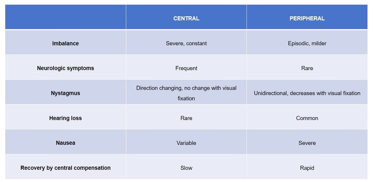
The imbalance is usually more severe in central causes, while peripheral causes tend to be episodic and milder.
Peripheral causes generally do not present with neurological symptoms. In peripheral cases, nystagmus is usually unidirectional, meaning the direction of the fast phase remains consistent. Hearing loss is uncommon in central causes. Nausea is usually more severe in peripheral causes. Recovery through central compensation is typically rapid in peripheral causes. Peripheral vertigo The duration of vertigo varies depending on the underlying condition:
Quick examination guide A quick examination guide involves performing an otoscopy to assess for conditions such as Otitis Media with perforation and Cholesteatoma. Cholesteatoma is one of the ear diseases that commonly causes acute vertigo. An active perforation on the left side with some crusting, or the presence of a large polyp in the ear of a patient experiencing vertigo or giddiness, warrants an ENT referral. Other important assessments include checking blood pressure, examining the pupils to determine whether they are equal and reactive, and looking for signs such as nystagmus and facial asymmetry. Additional examinations include pass-pointing and the head impulse test. Pass-pointing involves asking the patient to touch their nose and then touch the examiner’s finger. If the patient can accurately touch the tip of the examiner’s finger with their hand fully outstretched, it is unlikely to indicate a cerebellar or central nervous system issue. However, if the patient repeatedly misses the examiner’s finger despite having their arm fully outstretched, this may suggest a cerebellar problem. “The hand must be fully outstretched. It cannot be half extended. Missing your finger repeatedly with the hand fully outstretched could indicate a cerebellar issue.” The head impulse test is routinely performed in clinics and is also known as the Halmagyi test. Vestibulospinal tests Vestibular testing includes several assessments that evaluate how the balance system within the inner ear, known as the vestibular system, is functioning. These tests are sometimes referred to as a vestibular test battery. Hearing tests are a mandatory investigation for all cases of vertigo and tinnitus. Low-frequency hearing loss is frequently detected in Meniere’s disease. Radiology High-Resolution Computed Tomography (HRCT) is performed to assess infections and Cholesteatoma. Magnetic Resonance Imaging (MRI) is not routinely required for peripheral vestibular dysfunction. The purpose of these investigations is to localize the pathology causing vertigo through a process of elimination. Particle repositioning manoeuvres (PRM) Each particle repositioning maneuver is designed for a specific canal. For the lateral canal, a particular maneuver is performed. A different maneuver is used for the superior canal, and another for the anterior canal. A test is performed first to determine the location of the displaced particle. Based on the findings, a specific maneuver is then selected. The procedure is effective. However, it often needs to be performed three or more times for the particles to be completely cleared. This is because, in BPPV, it is often not just one large crystal that is moving. There may be one crystal together with sludge or smaller sandy particles that are also present. Tinnitus There are two main categories of tinnitus. The first is primary (idiopathic) tinnitus, where there is no identifiable cause. The other is secondary tinnitus, which is associated with underlying pathology, such as ear disease, medication-related causes or somatosensory tinnitus. Somatosensory tinnitus is associated with myofascial pain originating from trigger points, such as the temporomandibular joint (TMJ), occipital region and cervical spine. One of the key questions to ask is whether the tinnitus is unilateral. If it is unilateral, it is more likely to be peripheral in origin. Another important question is whether it is pulsatile. Pulsatile tinnitus is a red flag that warrants immediate referral, as it may indicate a vascular malformation or a glomus tumor. Fluctuating tinnitus is mainly seen in metabolic conditions. It is also important to ask about any history of neck injuries, dental treatment, temporomandibular joint problems, hearing loss and, of course, vertigo. Examination “If you need to determine whether a patient might have a temporomandibular joint issue, examine them. Ask them to open their mouth and look at their tongue. “If they have what is called a scalloped tongue, meaning indentations from the teeth along the edge of the tongue, they may have bruxism, which is teeth grinding. “If you examine the lower buccal mucosa and see a ridge that fits perfectly between the upper and lower teeth, that is also evidence of bruxism.” Patients may have bruxism without being aware of it because it commonly occurs during sleep. If their partner or spouse is present, ask whether they grind their teeth at night. Bruxism can contribute significantly to tinnitus and may increase the perception of what would otherwise be imperceptible tinnitus. “All of us actually have that baseline kind of tinnitus that we do not pay attention to.” The examination should include:
Particular attention should be paid to asymmetrical sensorineural hearing loss, which is considered a red flag. “One percent of patients presenting with tinnitus and high-frequency hearing loss have a Vestibular Schwannoma. Ten percent of patients with Vestibular Schwannoma present with tinnitus and unilateral high-frequency hearing loss. An MRI is performed for unilateral sensorineural hearing loss associated with tinnitus. For pulsatile tinnitus, an MRA is performed," says Dr Shailendra Sivalingam. Treatment Treatment is dependent on the underlying cause. Idiopathic tinnitus presents a particular challenge because an identifiable cause cannot be found. Tinnitus retraining therapy is an umbrella term describing treatment methods that include cognitive behavioral therapy, masking therapy, psychological counselling and, in selected cases, antidepressants. Cognitive behavioral therapy helps patients understand their condition and recognize that, although tinnitus can be very debilitating, it is not life-threatening. Masking therapy has become increasingly accessible, with many smartphone applications available that provide white noise, brown noise, running water, rainfall and other background sounds to help reduce the perception of tinnitus. Psychological counselling and antidepressants may benefit patients who are particularly troubled by their symptoms, although not all patients require this level of intervention. For secondary tinnitus associated with hearing loss, the use of a hearing aid is recommended because the hearing aid itself acts as a masker for tinnitus. Treatment of the underlying pathology will often resolve the tinnitus. Myofascial therapy and acupuncture are recommended for patients with no other identifiable cause, particularly those with TMJ disorders and neck problems. Acupuncture may be beneficial because it improves blood flow, which is thought to contribute to relieving myofascial trigger point pain. Medication There are currently no clear definitive recommendations regarding medication for tinnitus. Evidence supporting medication is largely anecdotal, with very limited evidence from randomized controlled trials (RCTs). Ginkgo may be tried, as it acts as a vasodilator, provided the patient has no adverse reactions. Betahistine is not prescribed for tinnitus itself, but rather for the treatment of vertigo. Neuromodulation Neuromodulation is a newer treatment approach that has emerged in recent years. It involves bimodal neuromodulation, combining electrical stimulation of the tongue with sound stimulation. The Lenire device became available in the United States in 2023. It promotes neuroplasticity to alter the sensitivity to, and attention paid to, tinnitus. Basically, you place a small clip on your tongue, which is paired with the device and headphones. You listen to sounds while, at the same time, the clip delivers gentle electrical impulses to your tongue. What you are trying to do is condition your brain by pairing sound with electrical stimulation from the tongue. This forms the basis of neuromodulation. Studies have shown it to be effective. Meniere's disease and endolymphatic hydrops “You need to have that whole group of symptoms. That is what the American Association mainly categorizes as Meniere's disease — vertigo, fluctuating hearing loss and tinnitus. The hearing loss must also meet a certain threshold," explains Dr Shailendra Sivalingam. Endolymphatic hydrops is characterized by a sensation of fullness in the ear, a blocked feeling and some degree of hearing loss, but without vertigo. If the patient does not fulfill the full diagnostic criteria for Meniere's disease, they may instead have endolymphatic hydrops. Hydrops refers to excessive accumulation of endolymphatic fluid within the inner ear. In some patients, this fluid accumulation is related to fluid retention associated with a high-sodium diet. This forms the rationale for treatment with steroids and diuretics. |
EXPLORE FURTHER

|
Could your child be missing sounds?Recognizing hearing loss early can prevent lifelong speech and developmental challenges
|

|
Could your ‘allergies’ actually be something else entirely?Key differences between allergic and non-allergic rhinitis
|

|
Is my child's snoring normal, or could it signal something more serious?Understanding sleep-disordered breathing, warning signs, diagnosis and treatment options
|

|
When should I worry about voice changes?Persistent hoarseness may be more than temporary strain or a lingering cold
|